
Studio 20
Signal and Image Processing for Medical Ultrasound
Build a basic ultrasound system from the ground up
Six days of signal and image processing, ultrasound fundamentals, and applied AI — ending with your own working modules and a final presentation.

When
20–25 July 2026
Six full days · 10am–5pm

Where
Chanakya university Global Campus
Devanahalli, Bengaluru

You earn
2 Credits
Certificate of Completion

Open to
All Students
UG, PG, PhD across schools + external participants
What you’ll do
This course introduces signal and image processing methods in medical ultrasound imaging. It is built for engineering students working in biomedical, electrical, electronics, and computer science. You’ll learn what ultrasound is and how an ultrasound imaging system works, implement simple signal and image processing methods yourself, and apply basic AI to a segmentation problem in ultrasound imaging.
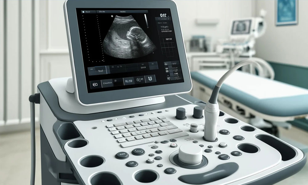
Ultrasound is a fundamental diagnostic imaging modality used in obstetrics, cardiology, radiology, and point-of-care imaging. It is portable, inexpensive, and can deliver immediate diagnosis to large populations. With the advent of on-edge AI, ultrasound is transforming medical care — providing immediate and affordable medical access at scale, and reducing the need for centralised expertise and patient care.
By the end of the studio, you should have the confidence to pursue jobs in the industry, or the interest to pursue higher studies in biomedical engineering.
What you’ll walk away with
By the end of the course, you will be able to:
- Understand an ultrasound imaging system and the concepts of system development
- Implement simple modules for basic signal and image processing in ultrasound
- Integrate code blocks to create a basic ultrasound system
- Apply AI in a practical application in medical imaging
- Work in teams and coordinate between teams to deliver goals
What you’ll make
- Code demonstrations
- A final presentation of your work
- A working prototype or application (where possible)
Your code, presentation, and prototype together form the assessment basis.
Your six days
| Mon | Introduction to signal and image processing (2 hours). Develop modules in MATLAB or Python to compute FFT, filter a signal, filter an image, and enhance contrast in an image (4 hours). |
| Tue | Introduction to ultrasound imaging (2 hours). Practical exposure to a hand-carried ultrasound system (4 hours). |
| Wed | Introduction to imaging modes (2 hours). Implement a basic B-mode imaging sequence (4 hours). |
| Thu | Introduction to Doppler ultrasound imaging (2 hours). Implement a basic Doppler imaging sequence (4 hours). |
| Fri | Introduction to signal and image enhancement methods (2 hours). Implement basic signal and image enhancements (4 hours). |
| Sat | Introduction to AI in ultrasound (2 hours). Object identification in ultrasound using AI; assessment and final presentation (4 hours). |
Sessions run full-day (approx. 10am–5pm). 2 hours of theory per day; 4 hours hands-on.
How you’ll be assessed
On four things: your ability to understand the concepts; your ability to apply the methods; your articulation of results; and the effort you put into the methods. Assessment will be a combination of these four.
Who it’s for
Engineering students in biomedical, electrical, electronics, or computer science. Prior familiarity with Python and/or MATLAB is recommended.
What you’ll need
Lab access will be provided — 5–6 PCs or workstations (preferably with Nvidia GPUs) running MATLAB, Python, Visual Studio Code, and PowerPoint, along with conference rooms for meetings, group discussions, and presentations. Bring your own laptop if you prefer working on it.
Your instructor
Dr. Seshadri Srinivasan
Dr. Seshadri Srinivasan is an internationally recognized imaging systems and ultrasound technology leader with over 25 years of cross-disciplinary engineering experience. He holds a Ph.D. in Electrical Engineering from the University of Houston and a Post Graduate credential in AI & Machine Learning Business Applications from the University of Texas, Austin. He currently serves as the Vice President of Imaging Systems at Exo Imaging Inc. in Santa Clara, California, where he leads the end-to-end development of next-generation, handheld MEMS-based ultrasound systems and open research platforms.
A prolific inventor and academic, he holds 35 patents (20 awarded) and has authored over 45 peer-reviewed journal articles and conference presentations, including foundational work in modulus imaging and ultrasound elastography. His studio provides engineering and biomedical science students with direct insight into signal and image processing pipelines, sensor integration, and on-edge AI applications used to build medical imaging systems from the ground up.

Registration open

Questions about a Studio?
Reach out to the Studios Coordinator. Happy to talk through any of the courses, what to expect day-to-day, or whether a particular Studio fits where you are in your learning right now.
Studios Coordinator
Anand K Sharma
cu.studios@chanakyauniversity.edu.in
+91 88930 33233
Campus
Chanakya University Global Campus
NH-648, Haraluru–Polanahalli
Near Kempegowda Intl. Airport
Devanahalli, Bengaluru — 562165